
The 200x Photo That Can Tell You If Your Hair Loss Is Reversible
Can Your Hair Follicles Still Grow Back? What a Simple Scalp Photo Can Reveal
Most people investigating hair loss have looked in the mirror a thousand times, searching their part line or hairline for evidence of what’s happening underneath. But the mirror is a blunt instrument. It shows you the aftermath of hair loss: thinner-looking hair, more visible scalp, without ever showing you the follicles themselves. That’s where the real story is written, and where the real answer to “can this still grow back?” actually lives.
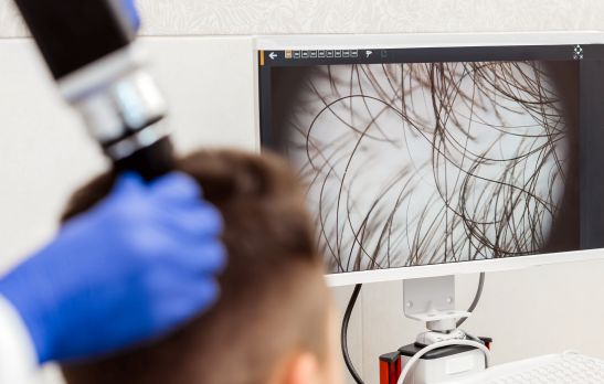
That is where the science of trichology comes in. Trichology is simply the branch of skin science dedicated to the hair and scalp. Think of it as dermatology’s specialty focused entirely on hair health. Trichologists, and dermatologists trained in this area, use a handheld magnifying camera called a trichoscope to photograph the scalp at up to 200 times its normal size. At that magnification, a trained eye can see individual hair follicles the way a specialist sees them: not as a general area of thinning, but as thousands of distinct, living (or no longer living) structures, each with its own story.
Quick answer: A magnified scalp exam uses a special camera to photograph individual hair follicles up close: their openings, the thickness of the hairs growing from them, and the skin surrounding them. It reveals whether each follicle is healthy, weakening, temporarily dormant, or permanently scarred. That’s information a mirror or a quick look at your part line simply cannot provide, and it directly shapes which hair restoration options will actually work for you.
This distinction, temporarily dormant versus permanently gone, is arguably the single most important piece of information in anyone’s hair loss journey. It’s the difference between “there’s still something here to work with” and “this particular follicle’s story is over.” Understanding how that determination is made, and what it looks like, can replace a lot of anxious guessing with something far more useful: realistic expectations.
What Is Trichology, and What Does the Camera Actually Do?
In plain terms, trichology is the study of hair: how it grows, why it stops growing, and what can be done about it. A trichoscope is simply the magnifying tool trichologists use to examine the scalp up close. It combines a digital camera, bright LED lighting, and magnifying lenses, typically in the 20x to 200x range, connected to a screen. That way, you can see exactly what the specialist sees, in real time, rather than taking their word for it.
At lower magnification (20x to 70x), the camera shows overall patterns across a section of scalp, including how densely packed the follicles are and how they’re arranged. At higher magnification (140x to 200x), individual hair shafts, the tiny openings hair grows out of, and the skin immediately around each one come into sharp focus.
This isn’t a gimmick. According to DermNet NZ, a widely respected dermatology resource, magnified scalp examination reveals specific, reproducible patterns tied to specific causes of hair loss. These patterns are simply invisible to the naked eye, but they help tell the difference between hair loss that’s likely to respond to treatment and hair loss that, unfortunately, is permanent.
What Shows Up in the Image that Your Mirror Can’t Show You
Here’s what becomes visible at high magnification that simply cannot be assessed by looking in a mirror or running your fingers through your hair.
- Hair shaft miniaturization. In androgenetic (pattern) hair loss, follicles don’t usually die suddenly. They shrink gradually over successive growth cycles. Under magnification, this shows up as a mix of hair shaft diameters within the same area: some full, healthy-caliber hairs next to visibly thinner, wispy ones. A systematic review published on PubMed Central describes this variation in shaft thickness, sometimes called anisotrichosis, as one of the clearest early signatures of pattern hair loss, often visible under a trichoscope well before thinning is obvious to the naked eye.
- Empty or reduced follicular units. A healthy scalp typically grows hair in follicular units containing two to four hair shafts. Under magnification, a trichologist can count how many units in a given area have their full complement of hairs versus how many have dropped to one hair, or show an empty follicular opening with no hair shaft present at all. The ratio of full units to empty or single-hair units is a meaningful density indicator that a general glance at the scalp will miss entirely.
- Perifollicular hyperkeratosis and follicular openings. This is a mouthful, but the concept is simple. It refers to a buildup of keratin (skin cell material) around the opening of a follicle, sometimes visible as a scaly collar or plug at the base of a hair shaft. Its presence, and whether the follicular opening itself is still visible and open, gives important clues about scalp health and inflammation. Research published in Actas Dermo-Sifiliográficas notes that whether a follicular opening is still present at all, as opposed to having disappeared entirely and been replaced by smooth, featureless skin, is one of the key ways a magnified exam distinguishes recoverable hair loss from permanent, scarring hair loss.
- The peripilar sign. A subtle brownish halo around a follicular opening, corresponding to mild inflammation beneath the surface. It’s commonly observed in pattern hair loss and, while not alarming on its own, is another data point in building a full picture of a follicle’s current state.
- Vellus versus terminal hairs. Under magnification, it’s easy to see the difference between vellus hairs (short, fine, unpigmented “peach fuzz”) and terminal hairs (the thicker, pigmented hairs that make up a normal head of hair). A rising proportion of vellus hair in an area that used to grow terminal hair is a visual record of miniaturization in progress.
None of these signs are visible with the naked eye. That is precisely why a magnified scalp analysis is considered a genuine diagnostic step, rather than a more dramatic way of confirming what someone already suspected.
Dormant and Savable, or Scarred and Inactive? A Simple Guide
This is the question underneath almost every other question a person has about their hair loss: is there still something here worth trying to save? Under magnification, dormant follicles and permanently scarred follicles tend to look meaningfully different, and that difference has real implications for which treatments are realistic. Below is a general guide to what each tends to look like and what it typically means. (This is educational context, not a substitute for an in-person evaluation. Trichoscopic patterns can overlap, and only a hands-on exam can make an individual determination.)
Signs a Follicle May Still Be Dormant and Savable
- The follicular opening is still visible, even without a hair shaft present. This suggests the follicular structure beneath the surface may still be intact, and is often considered dormant or miniaturized rather than destroyed, potentially responsive to treatments that support follicle health and hair shaft growth.
- There’s a mix of thick (terminal) and thin (miniaturized) hair shafts in the same area. This points to progressive miniaturization, typically consistent with pattern hair loss, and is frequently an early-to-moderate stage where intervention has the most to work with.
- A rising number of fine vellus hairs are replacing terminal hairs. This indicates follicles cycling into a weakened growth pattern, but they are often still active, though producing progressively finer hair.
Signs a Follicle Is Likely Scarred and Inactive
- The follicular opening is no longer visible, with smooth, featureless skin in its place. This means the follicle has likely been replaced by scar tissue and is generally considered permanent. Hair will not regrow from that specific follicle.
- There’s perifollicular scaling, redness, or hyperkeratosis, along with the loss of surrounding follicular openings. This is consistent with an active or resolved inflammatory or scarring process. The underlying cause should be identified, since scarring can be progressive if left untreated.
The single most important distinction on this list is the presence or absence of a follicular opening. A follicle that has miniaturized still has a door, meaning the structure is present, even if what’s coming through it is thin or intermittent. A follicle that has scarred over has had that door permanently closed. Everything about a hair restoration plan, whether non-surgical therapies make sense, whether transplantation is realistic, or whether a replacement system is the more practical path, flows from understanding which category a person’s follicles fall into, and in what proportion.
Why This Distinction Changes the Conversation
Most people arrive at hair loss research assuming the question is “how much hair have I lost?” A trichological analysis reframes it into a more useful question: how much of what’s left is still alive, and what is it still capable of doing?
That reframing matters emotionally as much as clinically. Someone who sees miniaturizing-but-present follicles under magnification is looking at evidence that their hair loss, while real and worth addressing, has not closed every door. That’s a meaningfully different starting point than staring at a mirror and assuming the worst.
It also matters practically, because it shapes which category of solution actually fits the situation:
- Areas with predominantly miniaturized, dormant follicles are often the best candidates for therapies designed to support and strengthen existing follicles, such as Enhanced Plasma Therapy, which is specifically intended to improve the caliber and thickness of hair that hasn’t yet been lost entirely.
- Areas with confirmed follicular loss, where donor hair is available elsewhere on the scalp, may be candidates for surgical hair transplantation, which relocates living follicles rather than attempting to revive ones that are gone.
- Areas where scarring or advanced loss make regrowth unrealistic are often better served by expertly customized non-surgical hair replacement or women’s hair replacement systems, which restore appearance and density without depending on dormant follicles waking back up.
No single approach is universally better. The right one depends entirely on what the follicles themselves show, which is exactly why the analysis comes before the recommendation, not the other way around.
What to Expect During a Scalp Analysis
For anyone considering having this done, the process itself is straightforward and non-invasive. A practitioner typically sections the scalp and examines several representative areas, usually including both a stable reference zone (like the back of the scalp, which is less hormonally sensitive and often used as a baseline) and the areas of concern. The trichoscope’s camera feeds to a screen in real time, so the patient can see the same magnified images the practitioner is interpreting, rather than taking someone’s word for it.
There’s no discomfort involved. It’s essentially a specialized photograph, not a procedure. What it produces is a documented, visual baseline that can also be used later to track whether a treatment plan is actually working, by comparing follicular density and hair shaft diameter over time rather than relying on memory or guesswork.
Frequently Asked Questions
What happens during a trichological scalp analysis (a magnified scalp exam)?
A trained specialist uses a handheld digital camera (a trichoscope) to photograph the scalp at magnifications typically ranging from 20x to 200x. They look at how many follicles are still present and producing hair, how thick or thin the hairs are, and the condition of the skin immediately around each follicle. The exam is non-invasive, generally takes only a few minutes, and you can view the same magnified images the specialist sees, in real time.
How can you tell if a hair follicle is dormant or dead?
The clearest visual indicator is whether the follicular opening is still present. A dormant or miniaturized follicle typically still shows a visible follicular opening, even if it’s currently producing a thin hair shaft or no hair shaft at all. The underlying structure is intact. A follicle that has been replaced by scar tissue no longer shows a follicular opening at all; the skin in that spot appears smooth and featureless. This distinction is difficult to see with the naked eye but is usually clear under magnification.
What does perifollicular hyperkeratosis mean?
It refers to a buildup of keratin (skin cell material) around the base of a hair follicle, often visible as scaling or a plug-like collar at the follicular opening. It is a sign worth investigating further, since it can be associated with inflammatory conditions affecting the scalp. Its presence, and whether it’s accompanied by other signs like redness or loss of follicular openings, helps a specialist determine an underlying cause.
Can miniaturized hair follicles be reversed?
Miniaturization exists on a spectrum, and outcomes vary by individual, cause, and how advanced the process is. In general, follicles that are miniaturized but still structurally present respond better to intervention than follicles that have already scarred over, which are considered permanent. This is exactly why identifying which stage a follicle is in matters more than treating hair loss as a single, uniform condition.
Does a magnified scalp exam hurt, or is it invasive?
No. It is essentially having a specialized close-up photograph taken. There’s no cutting, injecting, or discomfort involved. The camera simply magnifies and photographs the surface of your scalp.
If you have been looking in the mirror trying to guess what is really happening beneath your hairline, a magnified scalp exam replaces that guesswork with an actual picture, one that shows which follicles are still in the game and which restoration options genuinely fit your situation. Our specialists at Van Scoy Hair Clinics have been helping people across Ohio understand exactly this, with honesty and without pressure, for over 50 years. Schedule your free, private consultation to see what a close-up look at your own scalp reveals.
Sources referenced for clinical accuracy: DermNet NZ, PubMed Central (National Library of Medicine), and Actas Dermo-Sifiliográficas.
